Improve The Definition & Intensity of a Wright’s or Wright-Giemsa Stain

Looking to improve the definition of your Wright’s or Wright-Giemsa stain? We can help! Classical hematology stains are useful for clear structure differentiation within blood samples. However, there can be differences in how the stains are perceived by the pathologist you’re working with – she or he may want to see different staining intensity for certain types of cells or morphological components.
In addition, issues can occur within the procedure to cause undefined neutrophils, poor nuclear staining definition, or general feature staining that is too weak or undefined.
Our team of application experts understands these hematology staining issues and quickly makes recommendations to our clients. Here are their frequent recommendations when it comes to improving the definition and intensity of a Wright’s or Wright-Giemsa stain for hematology.
Solving for Weak or Poor Intensity Eosinophilic Staining
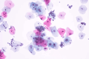
Since eosin provides the pink color to the cytoplasm within cells, weak eosinophilic staining causes the specimen features to blend together. This makes it difficult to differentiate between certain cell structures and/or red blood cells within the specimen. Often used in automated slide stainers or integrated systems, these Classical stains can require some specific adjustments to ensure the proper visual intensity in your specimens. There are typically three common issues we encounter. Here we’ll identify their causes and subsequent solutions.
- The specimen structure was degraded. If degradation of the specimen causes poor intensity due to a delay between fixation and staining, proceed by fixing a fresh smear and then move forward with your normal staining protocol. Adjust your procedure in the future to avoid this issue.
- The pH in the buffer needs to be more eosinophilic. Switching from a 7.2 pH buffer to 6.8 pH buffer will strengthen the buffer to be more eosinophilic, causing more of the intensity or distinction in the red blood cells and eosinophilic morphology.
- The buffer ratio has been compromised. Ensure that the buffer/stain mix ratio has not been altered by increased throughput or by extended time between uses. To be safe, swap the solution for a new mix. This should be done every six hours to ensure strong intensity.
Solving for Basophilic Staining is Too Weak or Has Poor Definition
Weak staining of white cell morphology can also be a symptom of issues with Wright or Wright-Giemsa stains, causing a lack of definition of key white cell components and/or interfering with cell differentiation. Try one of these solutions if the basophilic staining definition is poor when using a Wright or Wright-Giemsa stain:
- Reduce the time between fixation and staining. If the slide is not fixed quickly enough, the intensity of the stain will decrease.
- Change out the stain/buffer solution. The most important part of the procedure for a well-defined basophilic stain is the stain/buffer mix (usually 1:5 20% stain, 80% buffer). You might initially think to change out the 100% stain solution, but the actual staining takes place in the stain/buffer solution. If the stain/buffer solution has not been changed in over six hours, it’s time to change out the solution to ensure intensity in staining stays strong.
- You can also increase the concentration of your stain/buffer solution. Some labs prefer to use a 1:10 ratio (10% stain, 90% buffer). Increasing the ratio of stain to 1:5 (20% stain, 80% buffer) can often improve definition.
- Switch from 6.8 to 7.2 buffer. The stains and the buffer – in combination – make the definition between nuclei and cytoplasm more intense. Tweak the buffer combination based on what you want to see under each stain. If you’re looking for a more basophilic definition, choose a 7.2. If you’re not getting the definition you’re seeking, switch from a 6.8 pH to a 7.2 pH.
- If that is unsuccessful, we recommend switching from Wright’s stain to a Wright-Giemsa stain. The Wright-Giemsa stain is formulated to produce more intense basophilic/nuclear staining, so making this change will enhance the definition in the proper direction.
Astral Diagnostics is committed to quality. It’s why we offer on-staff experts in the fields of hematology, histology, cytology, and microbiology. We offer dozens of additional stains and more extensive knowledge.