Sample Stability: How Time, Temperature, and Handling Influence Hematology Results
Hematology testing relies on evaluating intact blood cells. Even small changes in cell morphology can influence interpretation, which is why...
3 min read
Reticulocyte counts and white blood cell differentials can sometimes present a familiar challenge in the hematology lab: analyzer results that do not fully match what appears under the microscope. Most technicians have experienced that moment when the analyzer and the microscope seem to disagree.
 When that happens, troubleshooting often follows a practical workflow, man, machine, material; a process of elimination approach that is used in laboratories to determine where potential causes are evaluated by first eliminating the element of human error, errors in machinery, or errors in material. Most labs follow the same troubleshooting logic: check the person, check the machine, check the reagents, and only after that start wondering if the sample itself is just having a very dramatic day.
When discrepancies arise, many laboratories will first review patient history and previous analyzer results to determine whether certain flags have been triggered consistently without corresponding findings on the blood smear. In some cases, underlying pathologic conditions can generate analyzer flags that are not readily reflected on the slide and may not immediately suggest an instrument issue. However, if similar unexplained flags are observed across multiple patients without supporting slide evidence, laboratories will often investigate analyzer performance. Elements such as historical error data, calibration history, quality control results, and hematology analyzer consumables are typically among the first checkpoints reviewed.
Incomplete mixing can introduce issues like microclots and fibrin that can alter blood cell counts later on, and those effects don’t disappear once a sample reaches staining. Other issues, like platelet clumping or uneven cellular distribution, can continue to influence the interpretation of the differential if they go undetected.
When a standard EDTA tube is used, anti-coagulant choices rarely cause inconsistent results. Delays before staining, however, can lead to a pale cell appearance because aging cells do not absorb dye as strongly, which may be mistaken for weak stain performance.
Much of the final staining appearance traces back to the preparation steps that occur before the slides are examined. In many labs, the staining process is so routine that it feels automatic, until something looks just slightly off. If there are any variations in timing or buffer composition, this can change how dyes bind to cellular structures, which can alter contrast, granule visibility, and background tone.
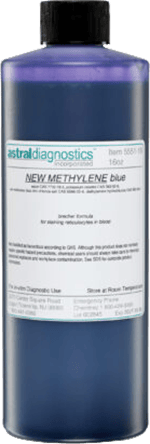 The Buffer-Stain Matrix
For supravital reticulocyte staining that uses new methylene blue (as well as differential stains) the buffer-stain mixture shapes the outcome long before microscopy. The ratio between the buffer and the stain affects color intensity, while pH stability will influence how eosin and basophilic components appear.
Different stains respond differently under these conditions. When peripheral blood smear stain precipitate is observed, there needs to be a review of buffer preparation and slide cleanliness to provide more insight before reagents are replaced immediately. More than one staining mystery has been solved by realizing the slide wasn’t as clean as everyone hoped it was. A reminder that sometimes the root cause isn’t complex—it’s just inconveniently simple.
Some staining patterns can resemble pathologic processes. For example, neutrophil granules appearing darker purple rather than a softer reddish tone may look like toxic granulation (overstaining or an imbalance within the buffer-stain mixture to create this appearance). Comparing multiple fields and preparation consistency can help differentiate an artifact from an inflammatory finding.
Flow cytometry flags help technicians identify where a smear requires a manual review. Automated counts highlight any potential abnormalities, and manual examinations can help confirm whether those findings reflect the morphology seen on the slide.
Discrepancies between analyzer results and smear appearance may stem from staining preparation or slide technique. If reagent replacement or preparation changes do not resolve the issue in the sample, then there has to be an evaluation of quality control trends that may reveal subtle shifts that have developed over time. Working methodically through operator technique, analyzer performance, and reagent preparation helps clarify whether a variation reflects process or pathology.
Laboratories striving for consistent reticulocyte and differential count accuracy benefit from dependable reagents and practical technical support. Ethos Biosciences provides high-quality stains, buffers, and diagnostic reagents designed for repeatable performance in routine hematology workflows. In addition to products, Ethos works directly with laboratory teams to help evaluate staining variables, investigate discrepancies, and support method consistency, helping labs maintain confidence in both automated results and manual slide interpretation.


Hematology testing relies on evaluating intact blood cells. Even small changes in cell morphology can influence interpretation, which is why...
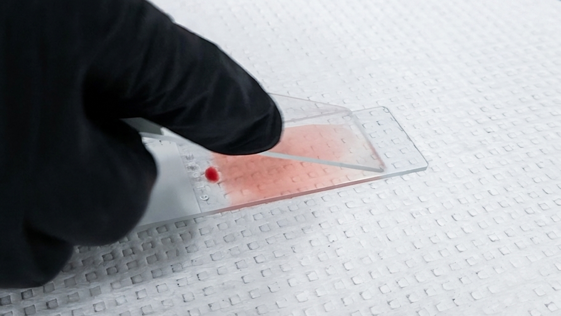
In hematology labs, accuracy and efficiency are critical. When processing blood smears, professionals need a staining solution that delivers...

Inconsistent white blood cell (WBC) or platelet counts are rarely due to the analyzer. More often, they trace back to events that occur before the...