Cytology workflows require careful control at every stage from initial specimen prep to staining and microscopic evaluation.
Ethos Biosciences provides a focused range of reagents and microscopy solutions that align with every stage of the process, from specimen collection through final analysis. Our reagents and stains help maintain consistent preparation and effective staining to provide dependable slide quality for both routine screening and more specialized applications.
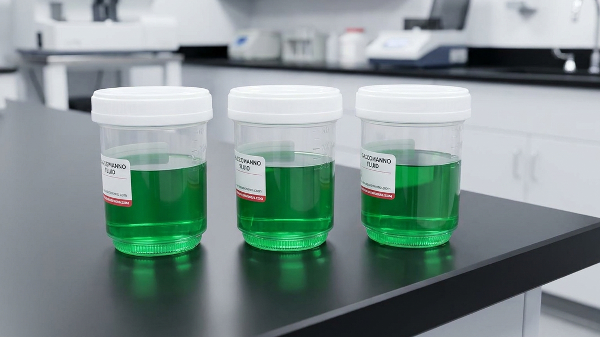
Sample collection and fixation work together to stabilize cellular structure before staining begins. Fixatives are used to preserve morphology and prepare cell membranes for dye uptake so that any staining needed further in the process reflects true cellular characteristics.
Routine stains are used to establish baseline cellular detail across a wide range of specimen types. These formulations highlight nuclear and cytoplasmic contrast, allowing abnormalities to be identified during initial review.
Hematological stains are adapted for cytology applications where blood or marrow elements are present. They bring out subtle differences in cell populations, which supports differentiation and a more detailed assessment.
In time-sensitive settings, rapid staining provides an early view of cellular details before full processing is complete. Quick stains are able to develop contrast within minutes, allowing samples to be assessed without delaying workflow.
After staining, dehydration, and the clearing steps, prepare slides for final mounting and long-term preservation. Controlled removal of water and replacement with clearing agents helps maintain clarity as well as prevent distortion during microscopy.
Routine stains can be modified or combined with other methods to emphasize specific features in specialty applications; an approach that builds on standard protocols without introducing any unnecessary complexity.
Mounting mediums protect stained specimens and maintain optical clarity over time and are used to create a stable interface between the slide and the coverslip, which helps preserve detail during repeated viewings.
The quality of the physical slide affects how evenly samples spread and how clearly they can be observed. Slides provide a clean, uniform surface that reduces visual interference and supports dependable examination across different sample types.
Immersion oil is used to improve the resolution of a slide at higher magnifications, as it helps reduce light refraction. Proper formulation supports a steadier optical performance and helps maintain image sharpness during analysis.
Strengthen Your Hematology Workflow from Collection to Diagnosis
Small inconsistencies in hematology can quickly lead to delays, uncertainty, and misinterpretation. This guide explores where procedural failures begin and how to correct them with practical, real-world insight.
Download the ebook to reduce variability, improve workflow reliability, and bring clarity back to every result.