1 min read
How to Achieve Reliable Blood Smear Staining in Seconds with Quick III™
In hematology labs, accuracy and efficiency are critical. When processing blood smears, professionals need a staining solution that delivers...
3 min read
Hematology testing relies on evaluating intact blood cells. Even small changes in cell morphology can influence interpretation, which is why stability from collection through analysis is so important.
In real laboratory workflows, however, samples do not always move perfectly from one step to the next. A tube may sit a little longer than expected, a courier run may take extra time, or a STAT sample may shift priorities in the middle of a busy shift. In most labs, the ideal workflow diagram looks perfectly linear. In reality, it often resembles a game of specimen Tetris.
When laboratories discuss sample stability, they are evaluating how reliably a specimen maintains its original cellular characteristics from collection through analysis. Even small deviations introduced during handling or storage can influence interpretation.
The elapsed time between sample collection and analysis can affect cellular structure, integrity, and analytical performance. Anyone who has worked a busy bench shift knows how quickly the clock can move. Between analyzer flags, incoming specimens, and routine workflow interruptions, it does not take much for processing delays to quietly accumulate.
 Most laboratories aim to process specimens as efficiently as possible, but real workflows are rarely perfectly linear. Each test has different timing requirements, but under standard hematology practice, EDTA-anticoagulated blood samples are typically analyzed within several hours of collection to preserve cellular integrity. This maintains the morphology needed for complete blood counts and differential analysis. If processing is delayed, progressive cellular degradation can occur, with neutrophils often exhibiting early structural degeneration and cytoplasmic vacuolization, along with increased platelet clumping. These changes can contribute to inaccurate results in automated systems, particularly affecting WBC differentials and platelet counts.
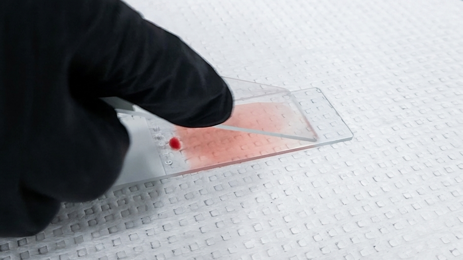
These changes are more frequent during microscopic smear review than during initial analysis. Labs that evaluate peripheral blood smear stain may see samples precipitate or may encounter irregular staining patterns, which may lead them to suspect reagent issues, but the overall elapsed time can be the problem or a contributing factor. When working with stains (like New Methylene Blue, Wright Stain and Giemsa Stain), the appearance of degraded cells can alter how the stain is absorbed and interpreted. Cell degradation can affect stain uptake and interpretation
Experienced laboratory professionals often develop an instinct for these subtle changes, recognizing when a sample simply looks “older” than expected under the microscope.
Subtle morphological findings often tell the story of what a sample has experienced before analysis. Poorly defined cell borders, uneven stain distribution, or irregular background staining can indicate that processing has been delayed.
When these findings are reviewed alongside analyzer data, laboratory professionals can often determine whether variability originates from sample age rather than problems with equipment, buffers, or staining reagents.
Temperature plays a huge part in the stability of samples. Elevated temperatures during storage or handling can accelerate degradation, in turn, increasing the fragility of the blood sample. This can lead to hemolysis.
 Additionally, cold exposure can also affect cellular morphology, leading to cell shrinkage or altered membrane appearance. All of which can resemble pathological changes when they are evaluated under a microscope. These temperature-driven effects highlight the need for consistent storage conditions throughout every stage of processing.
Biochemical variation may also occur when samples remain outside recommended temperature ranges. In an example, increased potassium values can sometimes be reflected in cellular breakdown rather than patient physiology. These patterns (and recognizing them) become particularly important when stains like Wright-Giemsa Fuccillo Stain are used to evaluate morphology alongside what the analyzer finds.
How samples are handled introduces another layer of variability that laboratories monitor every day. Mechanical stress from pneumatic transport systems or improper storage during transit can disrupt samples before they even reach the laboratory. Pneumatic tubes are great for speed, but from a cell’s perspective, it is less of a gentle ride and more of an amusement-park launch. Collection-related factors, such as inadequate mixing or expired collection tubes, may also introduce artifacts that resemble disease-related changes.
Because of this, troubleshooting stability issues often begins by reviewing workflow design and the path a sample takes from collection to analysis.
When these issues occur, troubleshooting will often begin by reviewing workflow design. The timing between collection, transit, and storage can be used to identify stability issues that influence staining performance or peripheral blood smear stain precipitate.
Reliable stains and reagents play an important role in evaluating sample stability. When laboratories can trust the consistency and performance of their stains, it becomes much easier to rule out reagent variability when unexpected findings appear. This allows teams to focus on other potential causes, such as sample stability or pre-analytical factors that may have influenced the specimen before analysis. High-quality products provide the confidence needed to distinguish whether a result reflects the true condition of the sample or a variable introduced earlier in the workflow.

1 min read
In hematology labs, accuracy and efficiency are critical. When processing blood smears, professionals need a staining solution that delivers...

1 min read
Hematology workflows begin long before stains are applied or slides are viewed under a microscope. When abnormal findings appear, laboratories have...
1 min read
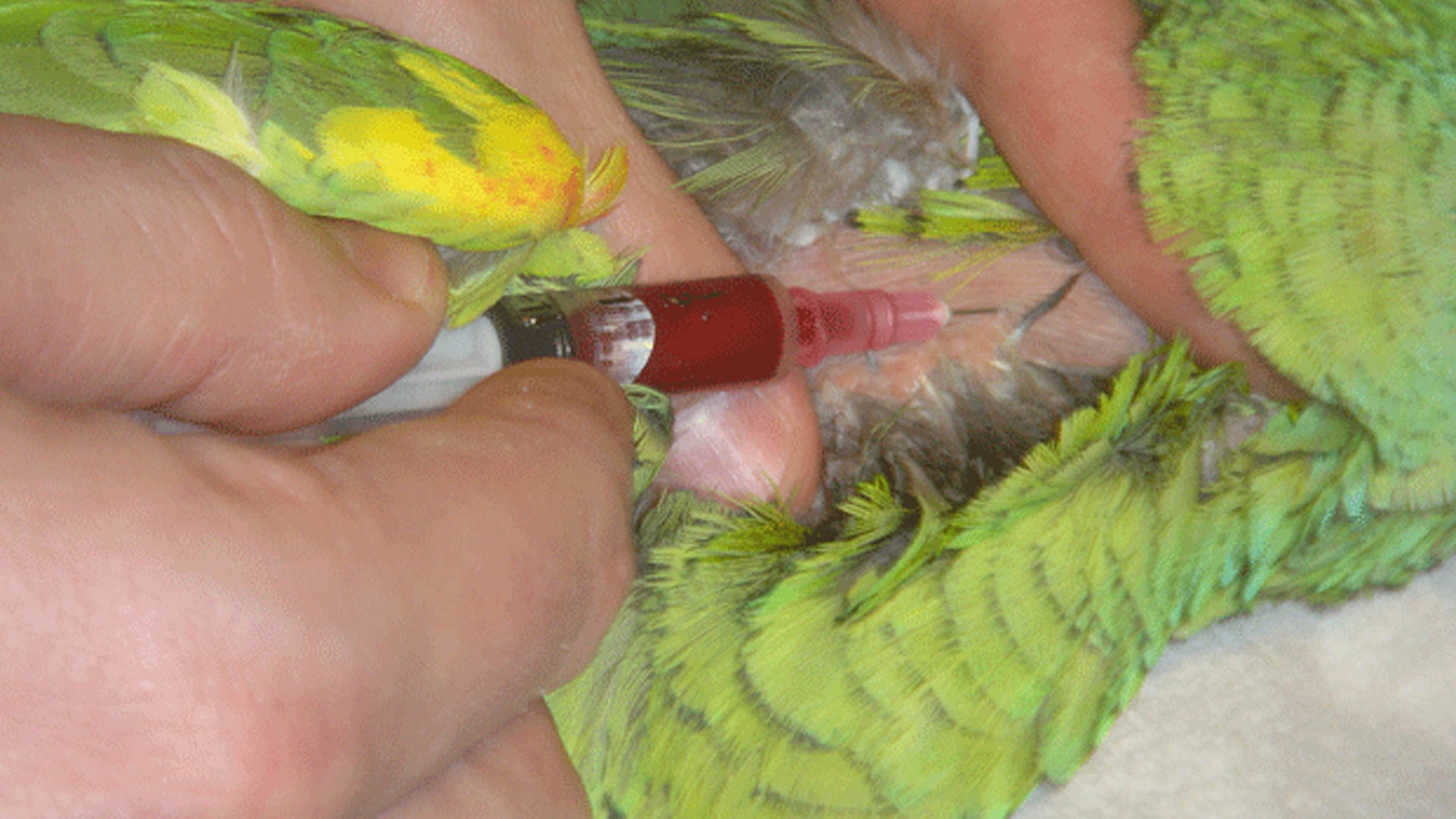
For veterinarians and animal scientists working with birds, reptiles, amphibians, and/or fish, obtaining accurate hematological data is vital for...