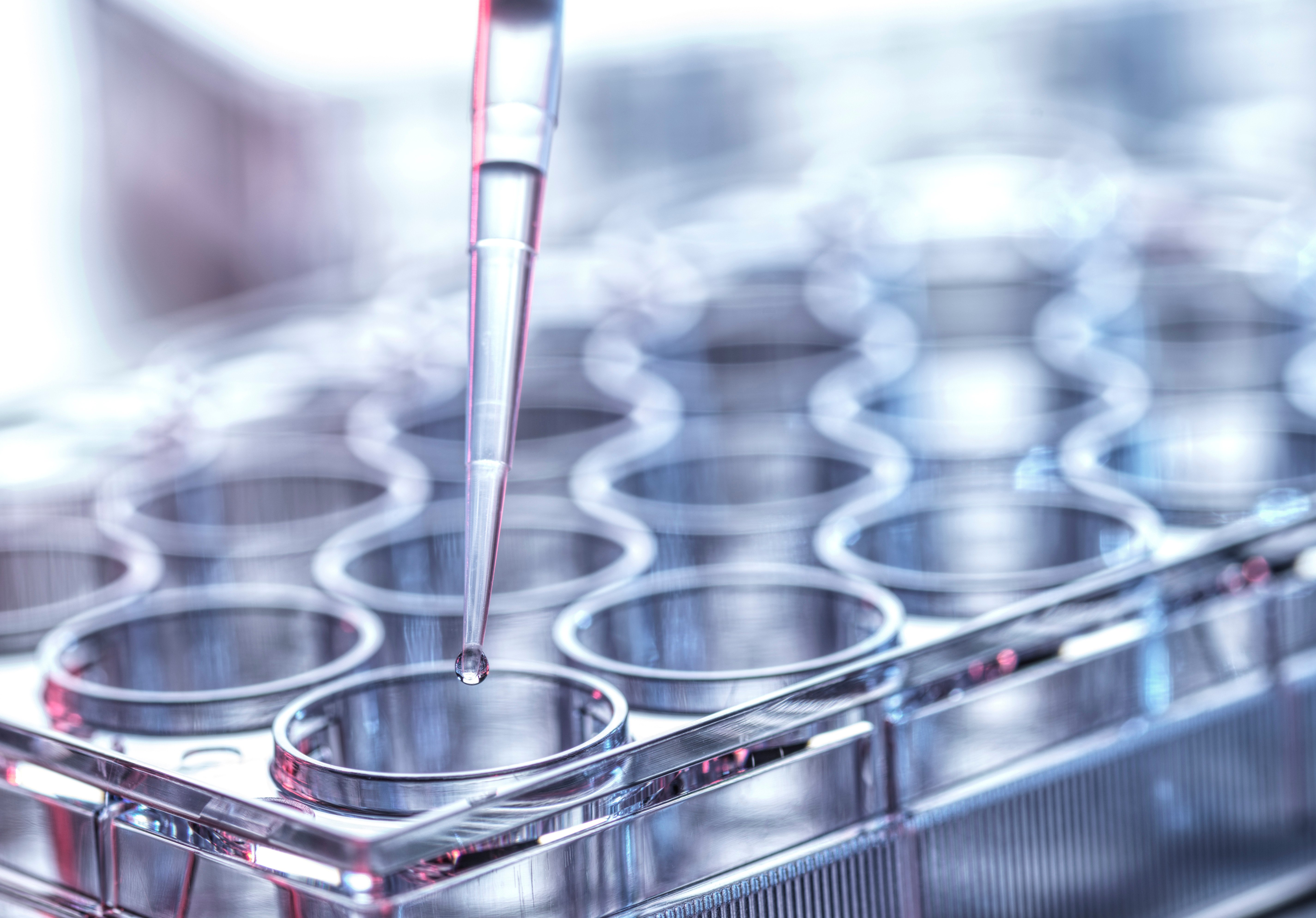
Microbiology laboratories follow a series of standardized steps to recover, identify, and evaluate microorganisms from clinical specimens. Ethos Biosciences supplies the materials needed to support each stage of that workflow, from specimen processing through organism identification and microscopic examination. Product selection at each step can influence how clearly microorganisms are recovered, differentiated, and viewed. The categories below are organized around the points in the workflow where those materials are used.
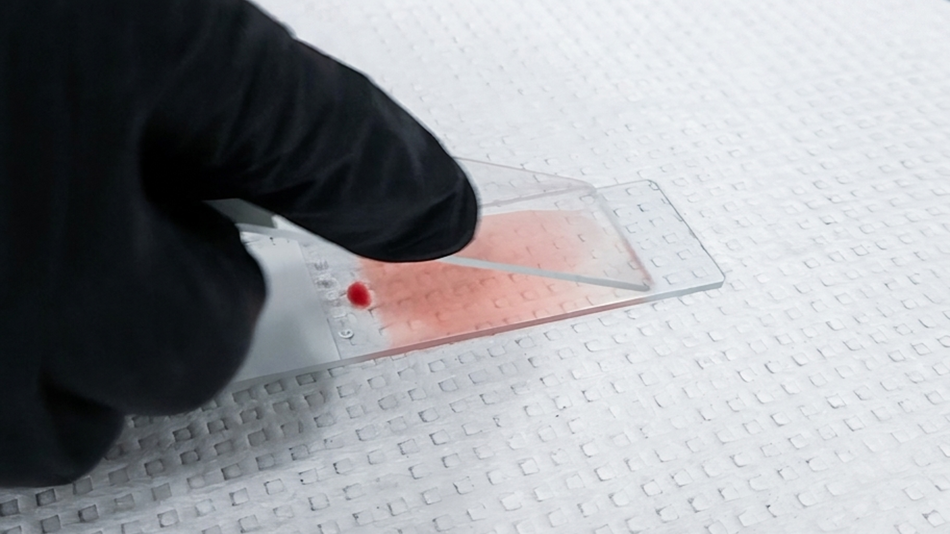
An early review of a specimen can reveal differences in staining behavior and cellular structure before culture results are available, as visual differences help laboratories assess smear findings. Then techs can decide what additional testing may be needed.
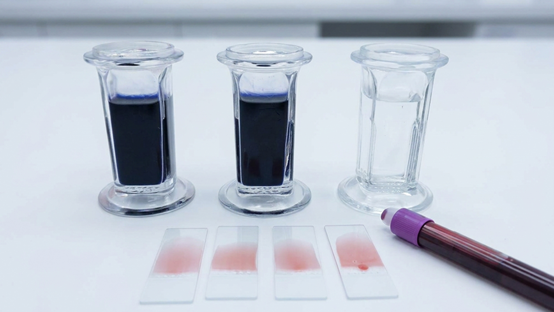
For commonly performed staining procedures, having the required reagents grouped into one kit can make day-to-day setup more straightforward. It also gives laboratories a more uniform process from one staining run to the next.
Certain bacteria have cell walls that do not respond well to conventional staining methods. Specialized acid-fast techniques make these organisms easier to detect and are commonly used when mycobacteria are suspected.
Laboratories that routinely perform mycobacterial screening often rely on acid-fast bacterial (AFB) kits to maintain organized staining. Bringing the required reagents together into one system helps simplify preparation and support a more uniform workflow.
Yeasts, molds, and other fungi may contain structures that are difficult to distinguish with general-purpose stains. Greater contrast makes those features easier to examine and can provide useful information during the initial evaluation of a specimen.
Parasitology stains enhance visualization of parasites and associated structures found in clinical specimens. These stains support the microscopic evaluation of protozoa, helminths, as well as other parasitic organisms.
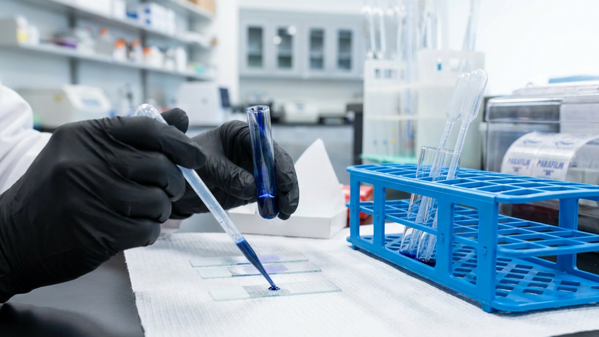
Once growth appears on a culture, staining can provide a closer look at the organisms selected for further workup. By judging differences in cell shape, arrangement, and staining behavior can help guide preliminary identification before additional testing is performed.
Microscopy mediums are used with prepared slides to support clear viewing beneath the coverslip and, depending on the formulation, the medium can also help preserve the specimen and reduce drying during storage.
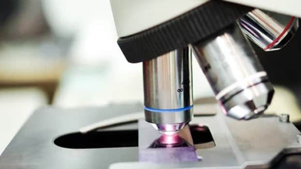
At higher magnification, oil placed between the slide and objective reduces the loss of light as it passes through the specimen. This creates a sharper image and makes fine microbial details easier to examine.
Strengthen Your Hematology Workflow from Collection to Diagnosis
Small inconsistencies in hematology can quickly lead to delays, uncertainty, and misinterpretation. This guide explores where procedural failures begin and how to correct them with practical, real-world insight.
Download the ebook to reduce variability, improve workflow reliability, and bring clarity back to every result.