Why Buffer pH Can Be Inconsistent and How It Affects Blood Smear Staining
Discover what causes buffer pH drift in hematology staining and how water quality, evaporation, storage, and contamination affect blood smear results.
2 min read
When a blood smear stain looks off, the first sign is often light staining. Cells may look pale, and nuclear detail may be harder to read. In other cases, the slide may lack the expected contrast. Other issues may show up as staining that looks too pink, too blue, or muddy instead of crisp.
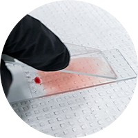 These changes can have several causes, including exhausted stain, carryover, stain oxidation, or poor handling. However, when the color balance shifts too red or too blue, buffer pH should be one of the first variables checked. In hematology staining, pH affects how dyes bind to cellular structures, which can change the appearance and readability of the smear.
The stain-buffer mixture is where much of the staining behavior takes place. In a typical hematology stain process, the straight stain step helps prepare the cells. The stain-buffer mixture allows the dyes to interact with cellular components. The final buffer rinse removes excess stain and precipitate.
Because this mixture controls much of the dye interaction, pH has a major influence on the final result. A buffer is not just a rinse or a substitute for water. It contains salts that help hold pH stable, allowing the staining chemistry to behave in a more predictable way.
When water is used instead of a buffer, that control is lost. Tap water can vary by location, and even DI water does not provide the same buffering capacity as a prepared solution.
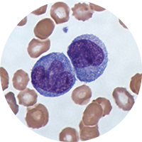 How well a blood smear is staining depends on the Romanowsky-Giemsa effect, or the effect that creates the reddish-purple nuclear detail and blue cytoplasm associated with well-stained hematology slides. This effect relies on the interaction between eosin, which contributes red or pink tones, and azure/methylene blue dyes, which contribute blue tones.
At a pH around 6.8, those dye interactions are balanced. The red and blue components can bind in the right proportion, helping produce the desired purple nuclear tone. If pH moves too far in either direction, one side of the staining reaction begins to dominate.
When the buffer used is too acidic, the smear may look too red or too pink. This is often described as more eosinophilic. When the buffer is too alkaline, the smear may shift too blue, or more basophilic.
With that in mind, some pH shifts can be intentional. For example, in parasitology, a slightly higher pH may create a more basophilic stain that makes parasite nuclei easier to see. In routine staining, though, an unexpected red or blue shift usually points back to the buffer.
If a pH issue is suspected, lab technicians should test the buffer with a pH meter, probe, or litmus paper and, if testing is not available, opening a fresh container may help rule out a material issue, ideally from a different lot.
When a blood smear stain shifts too red, too blue, or loses contrast, buffer pH is one of the first variables to check. Because pH affects how Romanowsky dyes bind to cellular structures, even small changes can alter nuclear detail and overall readability. In Part 2, we’ll cover why buffer pH drifts, including the effects of preparation, evaporation, storage conditions, water quality, and microbial growth.
Ethos Biosciences develops hematology stains and reagents designed to support consistent stain uptake, clear cell differentiation, and reliable workflow performance. To learn more about hematology stains and reagents, click here: Hematology Solutions.

Discover what causes buffer pH drift in hematology staining and how water quality, evaporation, storage, and contamination affect blood smear results.
Learn how stain selection improves avian blood differential accuracy by helping distinguish nucleated blood cells for clearer, more reliable...
Hematology testing relies on evaluating intact blood cells. Even small changes in cell morphology can influence interpretation, which is why...