1 min read
Choosing the Right Leukocyte Counting Method in Non-Mammalian Vertebrates
For veterinarians and animal scientists working with birds, reptiles, amphibians, and/or fish, obtaining accurate hematological data is vital for...
3 min read
Hematology workflows begin long before stains are applied or slides are viewed under a microscope. When abnormal findings appear, laboratories have to determine whether the results reflect true patient pathology or are instead related to variability introduced during pre-analytical handling or procedural factors such as staining, reagents, or instrumentation. Because hematology testing depends on evaluating intact blood cells, pre-analytical factors frequently become the first area of investigation. Understanding how these variables influence results helps labs approach their hematology testing through structured quality control.
 Laboratory professionals know that even the most advanced analyzers cannot compensate for variability introduced before a sample ever reaches the instrument. That reality is part of the daily rhythm of hematology work: identifying patterns, asking the right questions, and occasionally solving what feels a bit like a scientific mystery. Some days the answer is straightforward. Other days it may require retracing each step of the workflow, sometimes while juggling multiple STAT samples and hoping the analyzer cooperates.
Variability introduces ambiguity because the same visual and/or analytical findings can arise from multiple causes. Hematology tests, overall, measure cell size, morphology, and numerical distribution (meaning that physical damage to cells may resemble disease-related abnormalities). During troubleshooting, technologists often have to determine where the variability originates. This approach reflects the reality that collection, transport, and environmental exposure absolutely influence specimen integrity before any analysis and interpretation can begin.
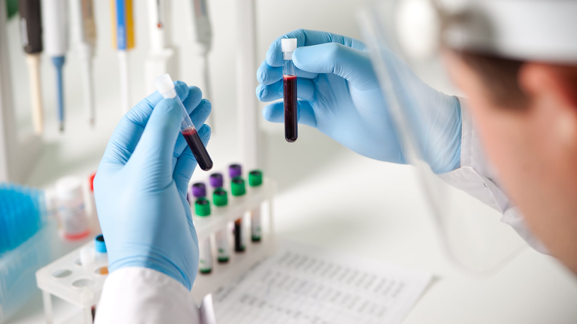
Collection techniques can introduce mechanical or chemical stressors that alter cells. Anticoagulants like K2EDTA help prevent clotting. However, inadequate mixing may produce microclots that distort cell counts or trigger abnormal flags within the analyzer. It’s one of those small steps that seems simple, until a perfectly avoidable microclot sends everyone on a fifteen-minute troubleshooting adventure.
Most laboratorians have encountered the occasional specimen that arrives looking less than ideal. While analyzers are remarkably sophisticated, they still rely on the integrity of the cells they evaluate. When collection variables interfere, the resulting data can send technologists down a troubleshooting path before the real work of interpretation even begins.
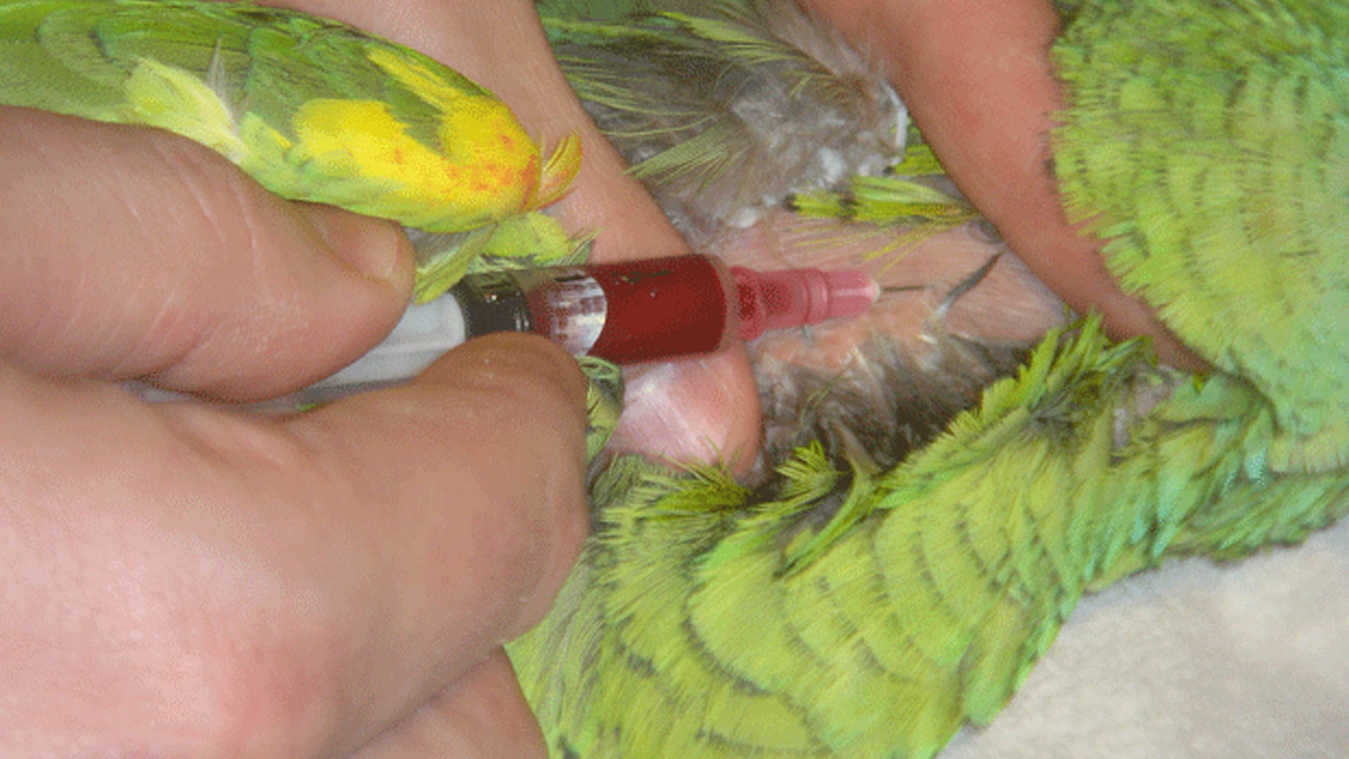
Collections taken from capillaries (via finger or heel sticks) may also show platelet clumping or other altered distribution patterns. Needle gauge also plays a role that can compromise results, as excessive vacuum pressure through narrow needles can damage red blood cells, introducing hemolysis that can further skew counts and downstream interpretation.
 Transportation and handling add another layer of variability that can impact hematology test results. Pneumatic tube systems and rough physical handling can affect cell membranes before a specimen reaches the lab. Sometimes a tube arrives looking like it experienced a slightly more enthusiastic ride than expected.
Heat can increase fragility, and cold exposure can change cellular appearance during a microscopic review. When analyzer results need additional investigation, they have to be evaluated to determine whether pre-analytical conditions explain the findings.
As samples move into analysis, abnormal results shift the workflow towards interpretation. Automated systems may flag results as irregular counts, which prompts slide preparation and review. During the reading of peripheral blood smears or the evaluation of stain precipitate, technologists have to look for patterns that differentiate specimen damage from true pathology. Burr cells, smudge cells, and platelet aggregation may suggest handling-related effects, while other findings can prompt a need for a deeper diagnostic review. Analyzer flags are often the first signal that something deserves a closer look, a reminder that even in highly automated laboratories, careful human review remains essential.
At this stage, reagents and stains are used to bring clarity to results. Stains help enhance visualizations so that morphology can be assessed with confidence, and the variations in color intensity or uneven staining may point toward procedural factors that are tied to stain preparation or reagent alcohol composition.
Consistent and reliable hematology workflows depend on predictable processes that allow labs to isolate the source of abnormal results. When reagents, dyes, and other hematology supplies perform consistently, technologists can focus on the origin of the abnormal findings rather than questioning the tools used to generate them.
Distinguishing between pre-analytical and procedural issues is a central part of hematology quality control. Careful attention to collection methods, specimen handling, and laboratory procedures helps ensure that abnormal findings reflect true patient conditions rather than avoidable variability. When each step of the workflow is consistent, laboratories can interpret hematology results with greater confidence and accuracy.

1 min read
For veterinarians and animal scientists working with birds, reptiles, amphibians, and/or fish, obtaining accurate hematological data is vital for...
1 min read
Hematology testing relies on evaluating intact blood cells. Even small changes in cell morphology can influence interpretation, which is why...

1 min read
Inconsistent white blood cell (WBC) or platelet counts are rarely due to the analyzer. More often, they trace back to events that occur before the...