1 min read
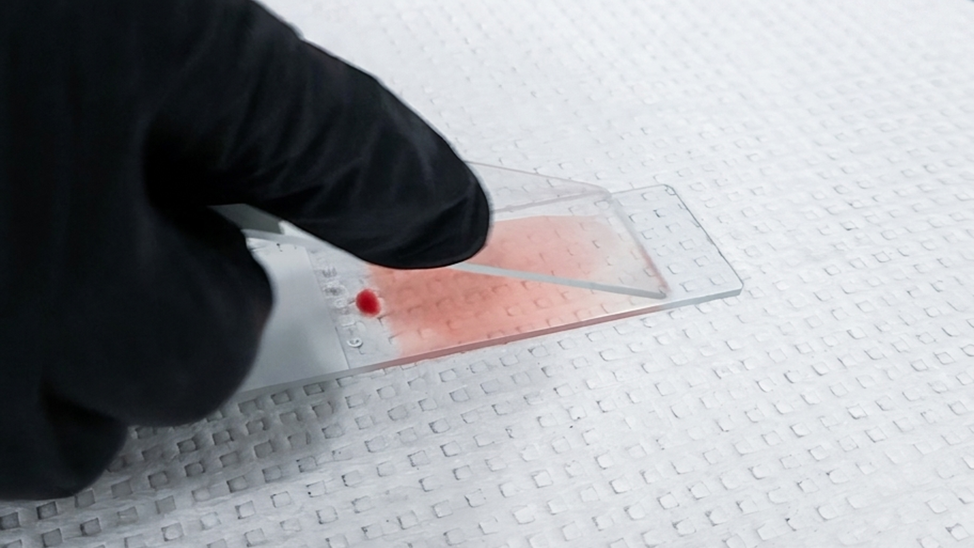
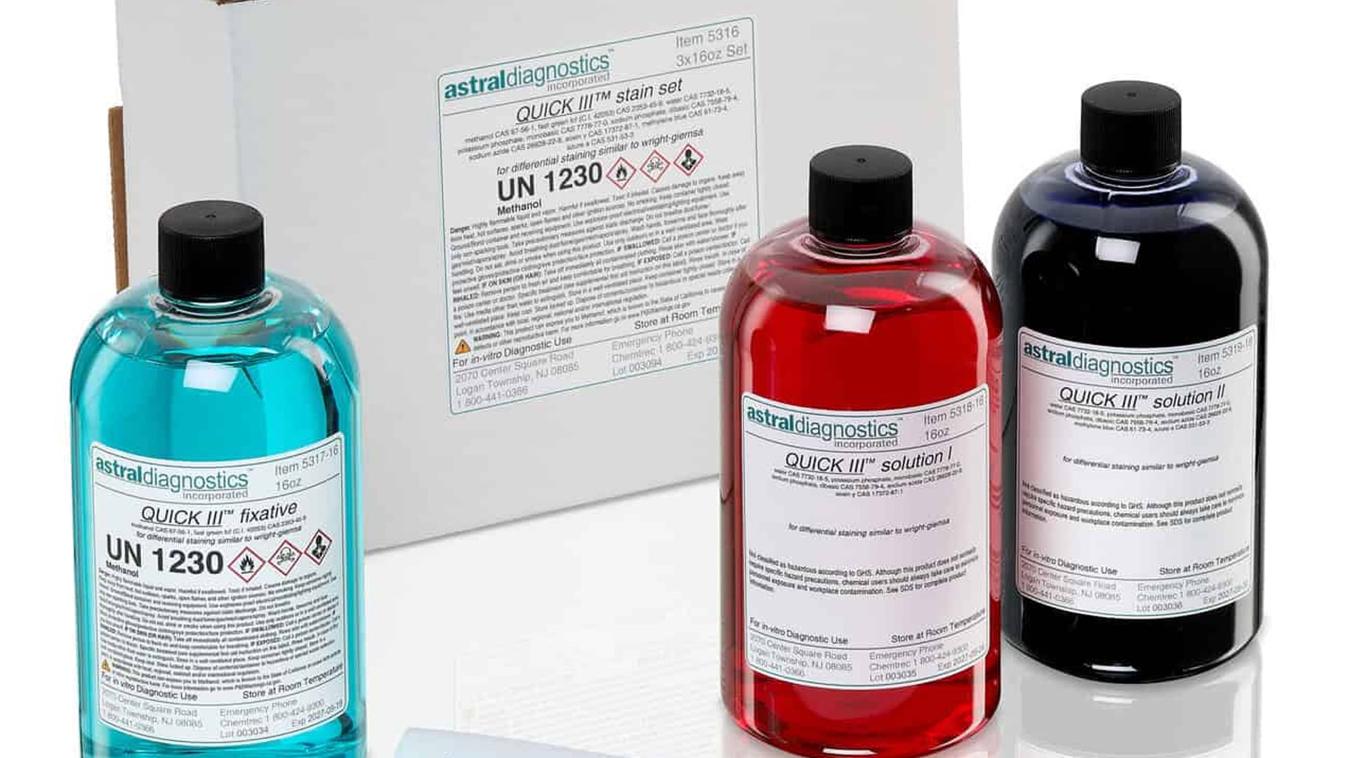
How to Achieve Reliable Blood Smear Staining in Seconds with Quick III™
In hematology labs, accuracy and efficiency are critical. When processing blood smears, professionals need a staining solution that delivers...
3 min read
For veterinarians and animal scientists working with birds, reptiles, amphibians, and/or fish, obtaining accurate hematological data is vital for diagnosing disease, monitoring immune response, and assessing overall health. A key part of this process is determining leukocyte (white blood cell) counts. Two commonly used techniques for these species are Phloxine B and Natt & Herrick’s Solution (NHS) (Natt & Herrick, 1952). Each has their own strengths and limitations.
Phloxine B is widely appreciated for its ease of use and precision (Gaunt, 2004). When used with a Neubauer ruled hemacytometer, it often yields more consistent WBC counts than Natt & Herrick technique (Dein et al., 1994). This makes it a practical choice for routine hematological evaluation in clinical or research settings.
However, this method does not measure the total WBCs directly. Phloxine B stains granulocytes, including heterophils, which are the avian equivalent to the mammalian neutrophil, and eosinophils, which function in immunity like their mammalian counterpart (Figure 1). This becomes a drawback when mononuclear leukocytes outnumber granulocytes, as it can skew results and reduce the accuracy of the total counts. This limitation should be kept in mind, particularly in species where mononuclear dominance is common (Campbell & Ellis, 2013).


Where more direct quantification of total leukocytes is necessary, especially when cell population is unpredictable, the Natt and Herrick’s method provides a more reliable approach. This technique involves diluting a blood sample at a 1:200 ratio with Natt and Herrick’s solution, which stains the leukocytes dark blue and allows for straightforward identification (Campbell & Ellis, 2013).
While this method may be slightly more time-consuming, the advantage of using this method is that a total erythrocyte (red blood cell) and thrombocyte count can also be obtained (Campbell & Ellis, 2013).
The following are suggested procedures for the Natt-Herricks technique (NHT) (Campbell & Ellis, 2013; Fudge, 2000):
Note: If there is difficulty differentiating between lymphocytes and thrombocytes, allow the sample to incubate in the Natt and Herrick’s for 60 minutes. This improves the differentiation between small lymphocytes and thrombocytes.
Campbell, TW, & Ellis, CK. (2013). Avian and exotic animal hematology and cytology. John Wiley & Sons.
Dein, FJ, Wilson, A, Fischer, D, et al. (1994). Avian leucocyte counting using the hemocytometer. Journal of Zoo and Wildlife Medicine, 432-437.
Fudge, AM. (2000). Avian complete blood count. Laboratory Medicine: Avian and Exotic Pets. Philadelphia, PA: WB Saunders, 16, 9-18.
Gaunt, SD. (2004). 9 – laboratory evaluation of leukocytes. In RL Cowell (Ed.), Veterinary clinical pathology secrets (pp. 33-37). Mosby. https://doi.org/https://doi.org/10.1016/B978-1-56053-633-8.50011-5
Natt, MP, & Herrick, CA. (1952). A new blood diluent for counting the erythrocytes and leucocytes of the chicken*. Poultry Science, 31(4), 735-738. https://doi.org/https://doi.org/10.3382/ps.0310735
Need more help? View our Resources page or contact our Technical Experts with any of your questions at: Info@ethosbiosciences.com or 1.800.441.0366. We’re always happy to help you!
1 min read
In hematology labs, accuracy and efficiency are critical. When processing blood smears, professionals need a staining solution that delivers...

1 min read
Nephrology focuses on the diagnosis and treatment of kidney conditions such as kidney function, kidney disease, and the preservation of kidney...
1 min read
Helicobacter pylori (H. pylori) is a spiral-shaped bacterium associated with chronic gastritis, peptic ulcers, and gastric cancer. Ethos Biosciences...