Ethos Biosciences develops stains and reagents for routine hematology testing, built around how samples move through smear prep and analyzers. Consistent stain uptake and clear separation of cell types allow technologists and lab technicians to interpret results without second-guessing if there is variation between slides.
Our portfolio includes Wright’s and Wright-Giemsa formulations, as well as supporting reagents, that maintain consistent staining behavior regardless of workflow. Products are manufactured using certified dyes and verified through specimen-based quality control. Our technical support is available to help laboratories adjust protocols and investigate performance issues when results fall outside the expected pattern.
Fixatives are used early in the process to hold cellular structure in place before any staining can begin. Without that stabilization, morphology can shift, which makes interpretation less reliable during differential review.
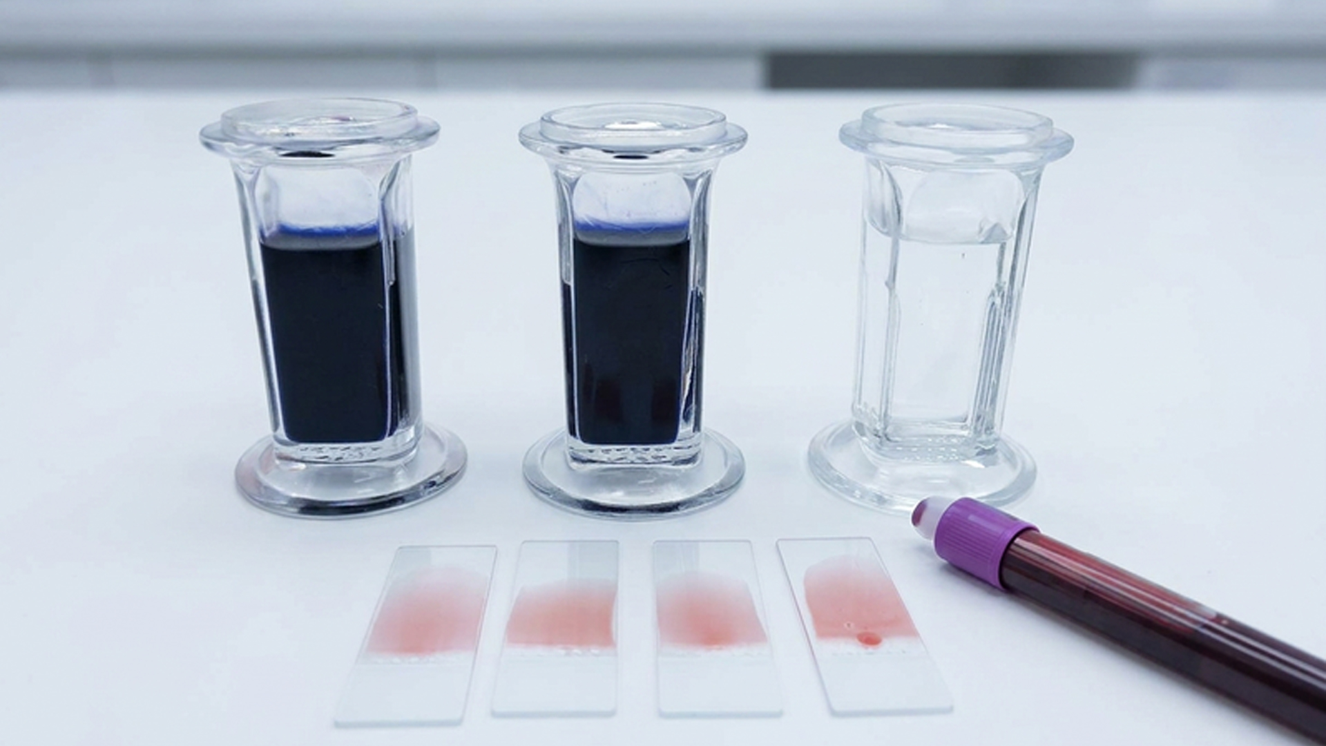
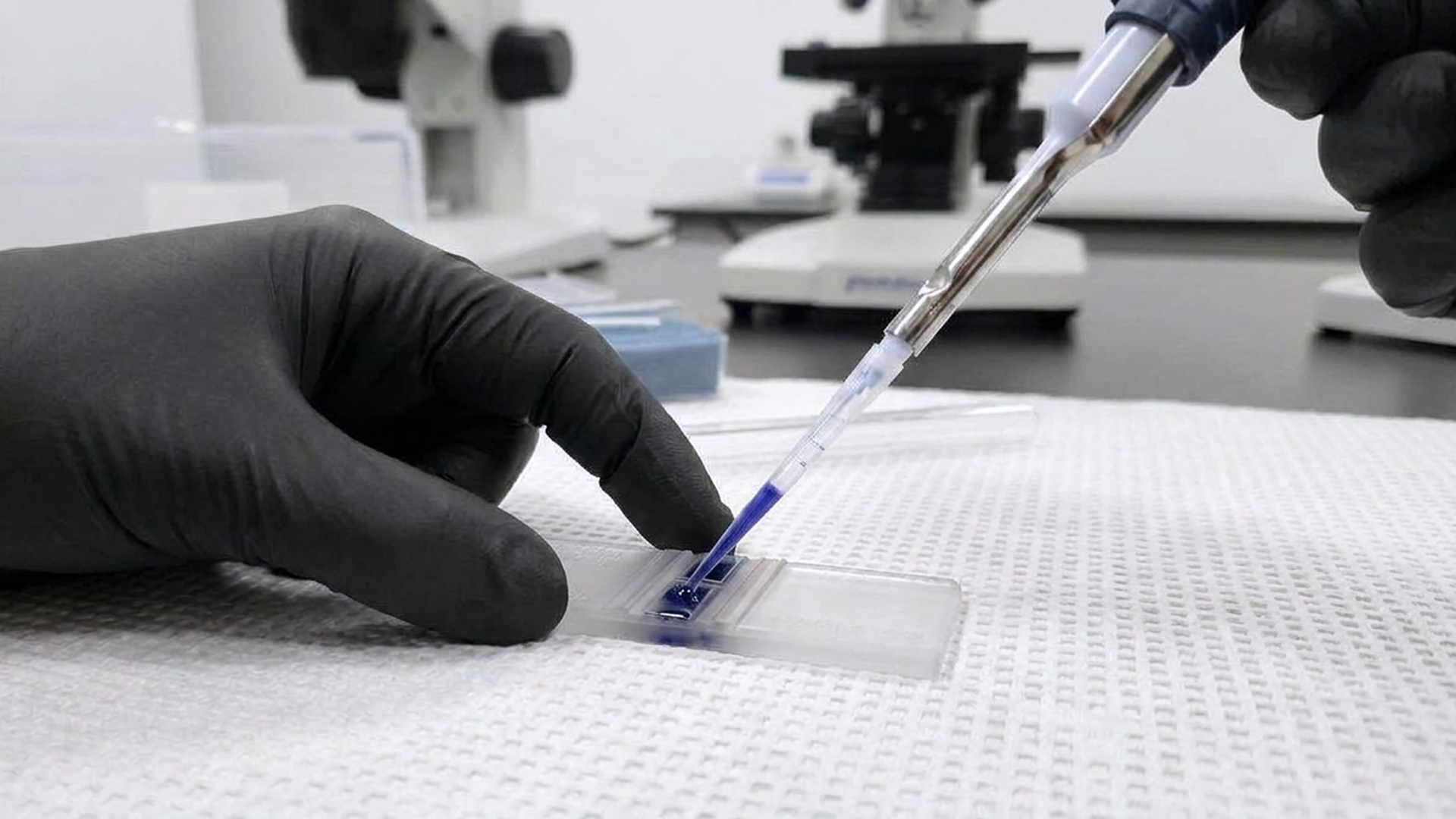
Sample concentration is adjusted with diluents so cell counts fall within a readable range. When dilution ratios are off, the distribution of the diluent becomes uneven, and that leads to results varying between runs and methods.
Red blood cells are ruptured from samples using lyse reagents to make white cell populations easier to see and count. This step clears the background and improves visibility for all types of workflows.
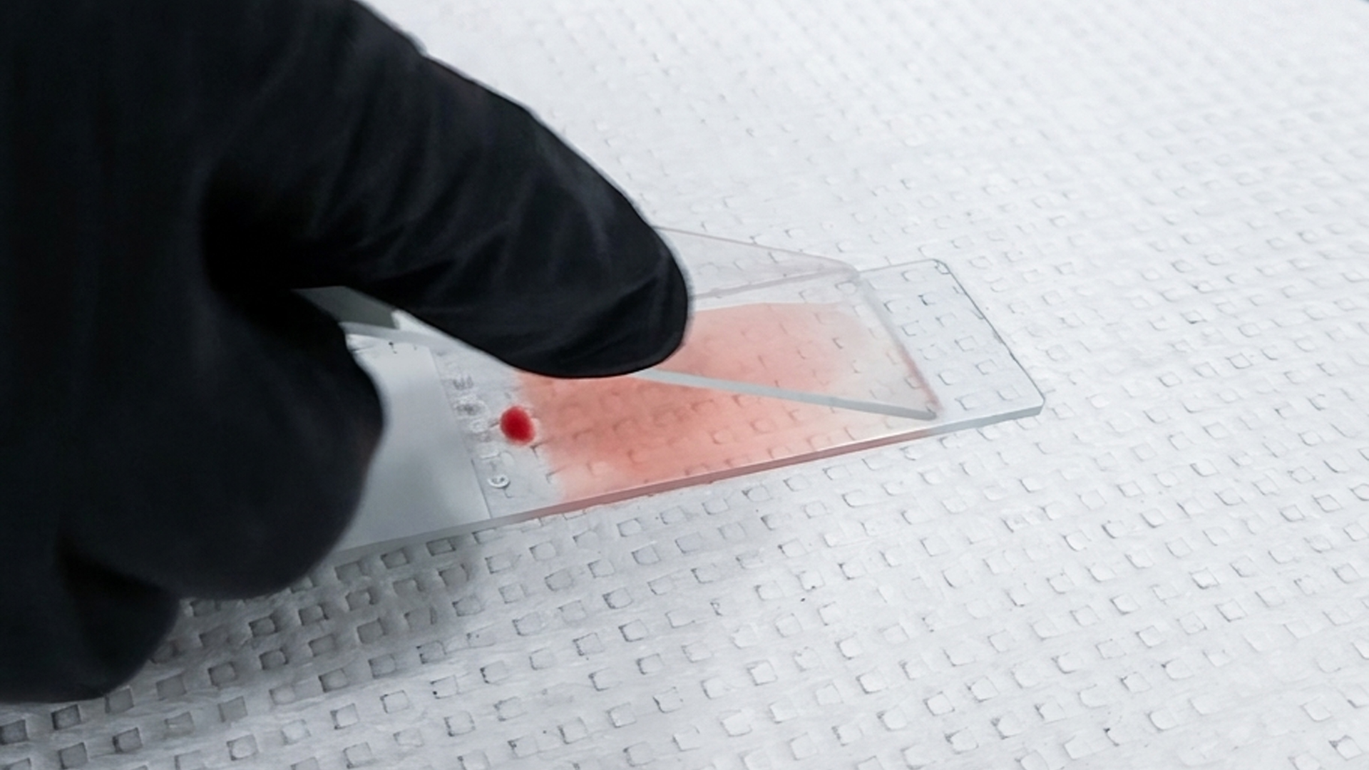
These stains are used to distinguish major blood cell populations during standard morphology review. Clear contrast between cell types helps support identification during daily differential work.
Formulations in this category are built for use on automated staining systems that run at higher volumes. Performance remains stable across batches, which helps limit variation when processing large numbers of samples.
Designed for shorter workflows, these stains allow slides to be prepared and reviewed in less time than in standard timelines. They are often used when results are needed quickly and still require readable cellular detail.
Staining outcomes depend heavily on pH conditions during processing. Proper buffering helps maintain color balance and keeps differentiation between cell types that are consistent from slide to slide.
Iron content can be visualized within cells to assess how it is stored and distributed. This is often used when evaluating suspected anemia or disorders tied to iron metabolism.
Applied to living cells, these stains make it possible to examine features that are not preserved after fixation. Reticulocyte counts and maturation patterns can be assessed more directly in this state.
Manual counting methods rely on improved contrast within the counting chamber to help separate cells more clearly. These stains support visibility between living and dead cells by staining the cytoplasm of dead cells, allowing living cells to be counted more easily.
Some findings require a more targeted structure, or some organisms do not respond to routine staining procedures. In those cases, more specific formulations are used to bring out details needed for further interpretation.
Sperm structure must be preserved before morphology can be evaluated under the microscope. Proper fixation helps maintain cell shapes and prevents distortion during slide preparation.
Preparation may involve dilution or buffering before analysis to bring cell counts into a workable range. These steps support accurate cell counting and help preserve motility characteristics during evaluation.
Visual contrast plays a larger role when examining sperm cells and their structures. Stains bring out details in the head, midpiece, and tail, highlighting abnormalities for review.
After staining, a mounting medium is applied to protect the sample and preserve any optical properties during an examination. This layer helps maintain clarity while allowing for limited changes that can occur over time.
Slide surface quality plays a role in how samples spread, adhere, and appear under the microscope. Well-prepared slides reduce background interference and help maintain clear viewing conditions during analysis.
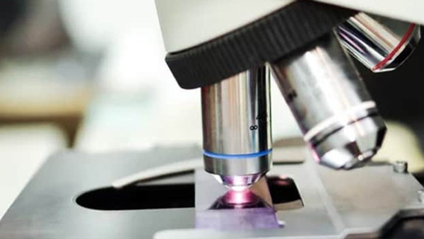
At higher magnifications, light refraction can limit what the objective captures. By using an immersion oil, the resolution of the image is improved by matching the optical paths, which allows finer cellular details to come into view.
Strengthen Your Hematology Workflow from Collection to Diagnosis
Small inconsistencies in hematology can quickly lead to delays, uncertainty, and misinterpretation. This guide explores where procedural failures begin and how to correct them with practical, real-world insight.
Download the ebook to reduce variability, improve workflow reliability, and bring clarity back to every result.