Choosing The Best Hematoxylin For Your Cytology Stain
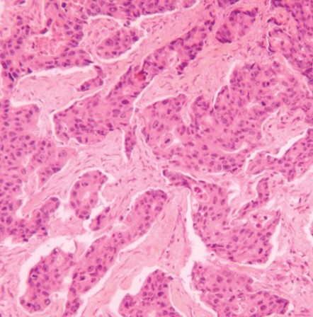
For cytology stains, you’re really selecting between one of three hematoxylin stains: Gill 1X hematoxylin, Gill 2X hematoxylin, and Harris Hematoxylin, Acidified. Here is how you can choose the best hematoxylin for your cytology stain, and the protocol for the two most common stains chosen by laboratory technicians.
Many choose based on the type of specimen being analyzed. Here, we’ve outlined the best use-cases for each hematoxylin.
Identifying cancerous cells, infections, inflammation, or other cellular abnormalities in cytology specimens
Generally, the progressive Gill’s Hematoxylin is best for evaluating cytology specimens. Most frequently, we recommend Gill 1X because it’s considered the lightest stain among the Gill Hematoxylin stains. It can complement the OG and EA stains without over-staining the specimens. Occasionally, we see that a Gill 2X Hematoxylin, such as when you’re analyzing cytology specimens for cancerous cells, infection, or other cellular abnormalities, is the right choice. This is largely dependent on your pathologist’s preference; if she or he prefers a darker purple stain, the Gill 2X Hematoxylin would be a better fit.
Harris Hematoxylin, Acidified is also commonly used for cytology stains and can be used progressively to yield acceptable results. This will yield a darker stain in comparison to Gill’s 1X Hematoxylin.
Find these hematoxylin stain protocols in our post, “How To Set-Up and Conduct H&E Stains.”
Gynecological Pap smear and non-gynecological cytological specimens
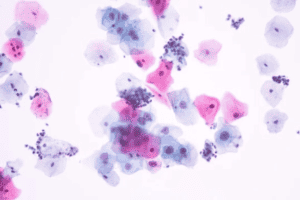
Cytological specimens, for example Pap smears or a bronchial wash/brush, generally benefit best from a Gill 1X Hematoxylin stain. This progressive stain is a light purple color that complements the OG and EA stains in the protocol without over-staining the specimen. Rarely is any stain more intense than 1X Gill Hematoxylin needed for this specimen type.
Protocol for Gill’s 1X and 2X Hematoxylin stain for cytology
- Fix cytological smears in 95% Ethanol: 15 minutes
- Rinse gently in DI water: 30 seconds
- Stain in hematoxylin solution, Gill 1X or Gill 2X: 1½ to 3 minutes
- Rinse gently in DI water: 1 minute
- Bluing Reagent: 15 to 60 seconds
- Rinse gently in DI water: 30 seconds
- Ethanol 95%:10 dips
- Papanicolaou Stain OG-6: 1 to 2 minutes
- Reagent Alcohol, 95%: 10 dips
- Papanicolaou Stain EA 50 or Papanicolaou Stain EA 65 EA: 2½ to 3 minutes
Note: the number indicates the proportion of the dyes. Use EA 50 for gynecological samples; and EA 65 for non-gynecological samples.
- Ethanol 95%, two changes: 10 dips each
- Ethanol 100%, two changes: 1 minute each
- Xylene or Xylene substitute, two changes: 1 minutes each
- Coverslip using Acrylic Mounting Medium, and examine under microscope
The progressive Harris Hematoxylin, Acidified protocol for cytology, as well as several others, are available in our ebook, Quick Reference Guide to Common Laboratory Biological Stains. Download it today!
If you’re working in a lab handling both cytology and histology specimens, then you may want to consider having more than one stain available: for example, Gill 1X Hematoxylin (for cytology) and Gill’s 3X Hematoxylin or a Harris (for histology). Determine your pathologist’s preference and choose the stain that best suits the need with these guidelines in mind.

