Choosing Between Wright’s Stains or Wright-Giemsa Stains
Choosing between Wright’s stains or Wright-Giemsa stains for your hematology specimens is often more a matter of your pathologist’s preference than about making a technically incorrect choice. That said, it’s important to know when these stains can be applied and when another option may be better.
Our ebook has a great decision-tree section to help you decide which of these two hematology stains to choose, depending on the type of specimen you’re testing, which equipment you have access to, and how much time you have available. The two Classic stains are Wright’s or Wright-Giemsa, and the other option is a Quick stain. Here is a breakdown by specimen type to help.
Testing: Blood morphology
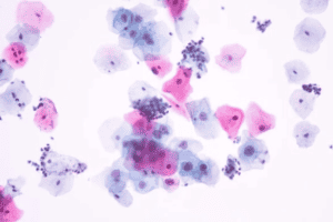
When conducting general blood cell analysis, immature red blood cell counts, immature white blood cell counts, platelet counts, or neutrophil counts, we recommend using either a Classic Wright or Wright-Giemsa stain. Most often, the basophilic nature of a Wright-Giemsa Stain gives the desired differentiation in white cell morphology that pathologists are looking for. The Wright-Giemsa stain is formulated to produce more intense basophilic/nuclear staining. If a more eosinophilic appearance is desired, choose the Wright’s stain instead.
If you’re looking for quick results or running a small number of samples, we recommend running a Quick Stain. This easy 3-step process will produce results similar to a Wright-Giemsa stain in under a minute.
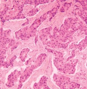
Testing: Bone marrow
Bone marrow specimens are typically assessed using a Wright-Giemsa stain to start. If the Wright-Giemsa is not distinct enough, we recommend using a Giemsa stain to improve stain intensity. For this specimen type, a Quick stain will not be effective since it is not formulated to penetrate bone marrow tissue within the standard staining time needed.
Testing: Allergens
Technicians will often test for Immunoglobulin E (IgE) antibodies using a Wright’s stain, since these antibodies bind to eosinophils in the cytoplasm of white blood cells resulting in a more intense, red color. A second option is a Quick stain protocol, since it, too, will provide the adequate red (eosin) staining required to identify IgE antibodies.
Testing: Bloodborne parasites
Testing for bloodborne parasites, including African trypanosomiasis, babesiosis, Chagas disease, and toxoplasmosis, is often best accomplished with a Wright-Giemsa stain. One notable disease differs from the rest; Giemsa stain is considered to be the standard stain for detection and identification of the malaria parasite. Quick stain can also be used in the detection of bloodborne parasites.
Protocols
The protocol for the Wright’s and Wright-Giemsa stains are the same. These stains follow the same protocol, with the second step being swapped for the preferred stain. The Wright-Giemsa stain is the most commonly used Classical stain by technicians because of its versatility and consistent staining appearance. It is formulated to produce more intense basophilic/nuclear staining in blood cell morphology. If you’d like to adjust to a more eosinophilic or less intense stain, select the Wright’s stain for your specimen. If you’re finding that your eosinophilic or basophilic staining is too strong, check out our post on adjusting Wright’s and Wright-Giemsa staining that’s too strong.
The Giemsa stain is used for assessing bone marrow specimens if the Wright-Giemsa stain is not distinct enough. It is also the main stain used for testing for malaria. This stain can help raise the intensity of the nuclear staining to be more basophilic. Its protocol is also the same as the Wright’s and Wright-Giemsa stains, except for the second step where Giemsa is swapped as the preferred stain.
We’ve designed a Biological Stain Decision Tree Map to assist lab technicians in choosing the best stain for the specimen they’re analyzing. Get it in our ebook, Quick Reference Guide to Common Laboratory Biological Stains. Whether your laboratory is using the Wright’s, Wright-Giemsa, or Quick stain for hematology, we want to see you succeed in identifying the best fit for your laboratory and specimens. If you didn’t find the answer here or need additional details, please don’t hesitate to reach out. We’re happy to help. Call one of our microscopy experts at 856-224-0900 or email at [email protected].